Tuesday, February 28, 2017

Perspective: Double Standards in Medicine


Image result for phone conversationIn a cold march morning in 2021 a big city urban “academic” hospital, the “consult” phone rings. A Nephrology attending answers “ Renal consult, can I help you?”.  The “Hospitalist” on the other end says, “ Yes, I have a creatinine of 1.5mg/dl, AKI, please see.”.  The Nephrologist replies, “ Yes, we shall, please send me details of the location and will take care of it.”  The Hospitalist says, “ thank you, and please don’t ask you fellow to see, I want an “Attending only” consult.  The Nephrology attending politely agrees.  He nods to the “renal fellow” next to him and says, “ Don’t worry, this one is for me only”. The Hospitalist continues “One more thing, I am done for the day, it’s 4PM, can you call the consult recommendations to my resident in house, thanks!” and hangs up.


Not too long ago, we trained as residents and fellows to see patients and get the “ nuts and bolts” of medicine and the field you were choosing. You wanted to see more patients to get the experience; perhaps not all of us but most of us wanted to get the “full and complete “ experience.  Attendings saw patients with us and we learnt from their clinical wisdom. While fellows/residents were work horses, most academic centers had educational missions as well to counterbalance the workload. Things have changed in the last decade. The above conversation reflects some of those changes.

What is wrong with the above conversation? What has bought us to this stage or might get us to this stage? Why is it “okay” for the “team” calling the consult to have trainees see their patients and “consult” team has to be “attending only”?  While a fellow might have less experience, their vision is not tunneled and they might bring an amazing differential diagnosis to the forefront.  While the fellow might be seeing many patients, seeing more patients might make them more efficient and learn to prioritize. There is lot of learning even when the volume of patients is high.  Is it the fear of “patient satisfaction” or is it a fear of “litigation”?  Not really sure.  I have heard it’s “communication” and many subspecialty fellows “don’t want to see more patients.”  Is that really true? Maybe once in a while, we all get tired and want to “go home”.  But we most of us went into medicine to “see patients” and provide optimal patient care.  I can say proudly that sometimes my patients ask “ Where is the fellow?, you are alone today? We miss the fellow..”  You form a team and a “team” always brings more to patient care than a “single person”. 

In addition, “consult” team cannot ask any questions. “ Yes sir, I shall see the patient”. Questions are asked to see the urgency of situation, to assess workup and to get a sense of what can be done quickly before we get there. “Asking questions on the phone” does not equate “avoiding” a consult. A good consultant will ask pertinent 1-2 questions and see the patient.  “Fellows” ask too many questions.. perhaps they are avoiding the consult.  Fellows ask questions to learn about the patient- it’s simple. Most fellows are nervous and want to make sure that when they present to their “attending”, they have a complete story.  Unfortunately, this is sometimes mistaken as “ avoiding consults”.  How quickly many attendings forget—“ I was once a trainee and did the same!”.
In the era of corporate medicine, where “academia” is blending with “private practice”, there is soon going to be no difference.  “Pan-consultemia” will drive these consults that will increase medical costs even more.   “Attending only” services are good to help off load the fellows burden in many academic centers and creating such services is an excellent idea for that reason.  When a surgical team calls a “attending only renal consult” and I don’t even get to speak to an “attending” on the other end-let alone a fellow or resident- it boggles my mind.


Double standards in medicine!

Friday, February 24, 2017

ASN Online Journal Chat on onconephrology topic

Dear Onco-Nephrology and Transplant Community members,

On Monday, February 27 at 9:00 pm EDT the Onco-Nephrology community is hosting a journal chat on immune checkpoint inhibitors and all ASN members are invited!

Here is how you can participate: 
  1. Join the Onco-Nephrology Community, if you haven't already
  2. Set your community notification settings for the Onco-Nephrology Community only to “Real Time”
  3. Read the Topic Summary on line or listed below.
  4. Watch your inbox on Monday for the discussion to start and participate in the chat
If you have any questions about setting up your notifications, please contact ASN Communities Associate Zach Cahill

Summary of topic by Mona Doshi, MD

The goal of any course of cancer treatment is to prevent and/or kill future growth of malignant cells. Sometimes this can be challenging as some cancer cells gain the ability to “trick” the immune system into thinking the cancer cells are normal healthy cells. Doctors are seeing promise in a group of drugs called immune checkpoint inhibitors, which actually “open up the immune system” and allow the immune system(T-cells) to recognize and attack the cancer. Two recent reviews published in early 2017 have summarized the effects of immune check point inhibitors (ICI) on the kidney.
We shall be discussing NEJM letter published on Jan 12th 2017. While effective in most cancer patients, this course of treatment has been less successful in kidney transplant patients because activating the immune system causes the patient’s body to start rejecting their donor kidney.  Five prior cases published in the literature of renal transplant patients getting PD-1 inhibitors have resulted in rejection. The rejections were mostly seen in PD-1 inhibitor based therapy compared to CTLA-4 therapy. In addition, the 2 cases of liver transplant where these agents were used and 1 case of heart transplant didn’t lead to a rejection episode.  But in the renal transplant patients, 5 cases have now been reported of leading to acute cellular and antibody mediated rejection when PD-1 inhibitor was administered. In a recent case correspondence in NEJM Jan 12th 2017 issue, the authors observed during the treatment of a patient living with cancer who had a kidney transplant that the combination of steroids and sirolimus (an immunosuppressant that has anti cancer properties),  could prevent a patient’s body from rejecting the organ during  cancer treatment with ICI.
In the case the authors observed the treatment of a 70 year-old Caucasian male who received a kidney transplant in 2010 and recently underwent treatment for small bowel cancer which had spread to the liver. The patient was given prednisone, a steroid, and sirolimus prior to incorporating an immune checkpoint inhibitor (nivolumab). The steroids were started 1 week prior to the starting of nivolumab and continued at a tapered regimen as mentioned in the manuscript to prevent the immune mediated reaction seen in prior cases. Steroids didn’t hinder the shrinkage of the cancer. There was significant response in tumor burden (as shown in the appendix) and the serum creatinine remained stable (as shown in appendix). There were no clinical or immunological signs of rejection.
In this forum discussion, as nephrologists, we can try to come up with ways to answer few questions for the oncologists.
1. What is the best treatment strategy for ICI induced AIN (dose, duration of steroids)?
2. What is the best preventive strategy for patients who have had ICI induced AIN and need to continue the targeted therapy?
3. Given the above single case report, can the above mTOR inhibitor+ steroid strategy be employed in all transplant patients receiving PD-1 inhibitors?


If you have questions about the content of the chat, contact any of the ONC leaders.
Sincerely,

The Onco-Nephrology Leadership Team

Wednesday, February 22, 2017

CONSULT ROUNDS: Anti-Phospholipid Syndrome and the Kidney

Renal abnormalities are present in approximately 7-9% of patients with primary anti phospholipid syndrome(PAPS).  Outcome and long-term follow-up usually are good. A large spectrum of renal thrombotic manifestations have been described in association with antiphospholipid antibodies, such as renal artery stenosis, renal infarction, renal vein thrombosis, acute or chronic thrombotic microangiopathy, and, more recently, the so-called “antiphospholipid antibodies nephropathy.”  Thrombosis can occur at any level of the renal vascular tree: Renal and intrarenal arteries, glomerular capillaries, and renal vein. Histologic findings show ischemic glomeruli and thrombotic lesions, without glomerular or arterial immune deposits on immunofluorescence. So besides TMA related findings, glomerular diseases can also be found with PAPS.
Other GNs that can be seen with PAPS is in this order:
Membranous GN
Proliferative GN
MCD-FSGS
C3 GN
The following two series in AJKD and CJASN suggest the varied degree of findings of glomerular and endothelial damage seen in the kidney with PAPS. 

In most patients, Lupus anticoagulant is positive (92.3%) along with b2 glycoprotein.
ANA is usually positive in low titer in >60% of patients whereas dsDNA might be only found in 30% of cases. Most patients have low complements levels( c3 and c4).

In the CJASN paper, all patients were treated with anticoagulation. Patients with MN were treated with steroids with addition of cyclophosphamide in two. Remission was achieved in two patients with MN; one was stable with mild proteinuria (0.5 g/d), and one patient was lost to follow-up. Both patients with diffuse proliferative glomerulonephritis were treated with corticosteroids plus cyclophosphamide with remission of nephrotic-range proteinuria. 

Eculizumab has been tried in one case report with PAPS associated TMA and another case with catastrophic PAPS.

Among them, renal involvement seems to be targeted in PAPS by other mechanisms such as immune complex deposition. Moreover, the heterogeneity of renal involvement confirms the presence of a continuum between SLE and PAPS  and suggests that a complete nephrologic workup should be performed in patients with signs of renal disease. Renal prognosis seems good if treated early as an autoimmune disease.


Clinical Case 89: Answers and Summary

Which is NOT a known renal complication of Bariatric surgery?


The renal risks of bariatric surgery are pre renal AKI, and long-term risks of nephrolithiasis and oxalate nephropathy. AKI is fairly common after bariatric surgery, with reports ranging from 2.9% to 8.5% in published studies, which have used varying definitions of AKI. Risk factors for AKI after bariatric surgery include higher BMI, lower eGFR, preoperative use of angiotensin-converting enzyme inhibitors, angiotensin receptor blockers, and intraoperative hypotension. Bariatric surgery patients may be prone to dehydration and higher risk for prerenal AKI in the long term, although this risk has not been quantified.  FSGS is a known secondary cause of Obesity but not related to bariartic surgery.

 A recent article in KI reports summarizes AKI related to bariatric surgery


Tuesday, February 21, 2017

In the NEWS: Death of Contrast induced Nephropathy

If you were a twitter follower: I am sure you saw this post by Dr Topf on


The article just published in Lancet proves in a RCT that fluids don't help in high risk patients in preventing contrast nephropathy (CIN).

http://www.sciencedirect.com/science/article/pii/S0140673617300570

This comes after many recent papers that have questioned the very existence of this entity. While it exists, perhaps the incidence has been over sized.  This was nicely shown by Chertow's team in 2016
https://www.ncbi.nlm.nih.gov/pubmed/27688297

Bigger questions will arise as we move forward in the era of Cardiac-Nephrology. Discussions with cardiologists are going to be essential. While the Lancet paper will get it's share of supporters and opposers, the debate will continue as now the question has arisen- does CIN even exist? Is this the death of CIN?


1. Does intra-arterial vs intra-venous contrast make the kidneys worse?
2. Does giving NAC matter?
3. Does giving IV fluids matter?- pre and post and or based on wedge pressure
4. Does holding ACEi/ARB matter?
5. Does holding diuretics matter?- Might so if they are planning to do a TAVR.

Tough road ahead as - what can the Nephrologist do then for these patients? I guess - Nothing! and perhaps doing nothing might be beneficial to the patient. We shall see!

Friday, February 17, 2017

Topic Discussion: Linear staining on IF and the differential diagnosis


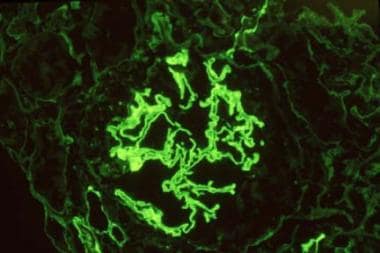
Linear Staining in IF on renal pathology differential diagnosis


Image result for linear staining IF


Classically the linear staining in taught in medical schools to be associated with anti GBM disease.

On a recent discussion on ASN-Communities on this topic by many glomerular experts led to generating a differential on other causes of IF linear staining when anti GBM serologies are negative.

Dr. Richard Glassock summarized the other causes on the forum as listed below with a few references:

1)    Atypical anti-GBM disease- This entity recently described by Nasr et al in 2016 is a more indolent form of Anti GBM disease where there are no serological markers for anti GBM and there is linear staining. It’s a slower disease and better renal outcomes compared to classic GBM disease. The light microscopy is variable from MPGN, TMA, to endocapillary proliferation and distinct lack of crescents. Some cases had deposits and some didn’t. 50% of these cases have a monoclonal disorder
2) Fibrillary GN with "pseudo-linear" IgG deposits, often secondary to autoimmunity, infection or cancer
3) IgG4 Anti-GBM disease- most assays do not detect IgG4 anti-GBM antibody 
5) Monoclonal IgG or IgA kappa directed to COLIValpha1/2 chains 
6) Wrong substrate, poor sensitivity, prozone phenomenon  in IF assays
7) "Immune sink" where all circulating antibody is bound to GBM sites in vivo-serial testing will often resolve this
8) Spontaneous decay of circulating antibody levels with persistence of tissue deposited antibody 
9) Diabetic Nephropathy: a physico-chemical alteration of GBM or IgG causing non-specific deposition of IgG (and albumin)


Other references


Saturday, February 4, 2017

Consult Rounds: Thrombocytopenia in ESRD


Thrombocytopenia in a hemodialysis patient can be from multiple reasons


Image result for thrombocytopenia


The top reasons we encounter are:

Medications
Medications
Medications

The most common is heparin and HIT.

A nice review of HIT in the renal patient is here

The other causes can be related to other medical illness not ESRD related. An important but rare cause that can be associated with dialysis associated with thrombocytopenia is a dialyzer reaction. A large systemic evaluation done in Canada suggested that the prevalence of thrombocytopenia associated with dialysis was due to the use of electron beam dialyzers( optiflux) rather than irradiated sterilized dialyzers ( others).  This suggests a relationship of use of the dialyzer sterilization technique causing this.  Exposure to e-beam radiation may change membrane integrity, structure, or physical properties of the dialyzer and could plausibly lead to platelet activation, aggregation, or adsorption and subsequent thrombocytopenia. E-beam sterilization has become a popular method of membrane sterilization recently, because it potentially allows a more focused and precisely delivered dose of sterilization toward the dialyzer membrane. A case report in AJKD had highlighted low platelets as a side effect of dialyzer reaction. Certain membranes due to the many types of hydroxyl groups contained can lead to an active complement system( the older dialyzers).  The degree of complement activation determines the biocompatibility of the membrane. A prior post in RFN also had discussed this phenomenon.
This review from Hemodialysis International is a great one for all to review.


If the patient is inpatient, we might notice that the day after dialysis the platelets drop from 150 to 50 perhaps and then rebound and then again the drop happens on the next set of labs. It is very likely that we are missing this phenomenon on the outpatient and there is ongoing endothelial damage happening chronically due to some of these membranes.  We only check monthly labs in US.  A true incidence of this phenomenon is hard to determine.  

Friday, February 3, 2017

Clinical Case 89: Answers and Summary

Which anti melanoma therapy can lead to immune mediated hyponatremia?


Ipilimumab(CTLA-4 inhibitor) has also been associated with electrolyte disturbances. Two cases of ipilimumab-induced hyponatremia due to panhypopituitarism from ipilimumab related hypophysitis have been reported. The incidence of hypophysitis in patients treated with this agent is close to 17% in clinical trials. Mechanistically, a loss of adrenocorticotrophic hormone-secreting corticotrophs leads to a secondary adrenal insufficiency and loss of regulatory effects of cortisol on arginine vasopressin. This could be the mechanism leading to the hyponatremia. While FDA reporting system has mentioned hyponatremia in PD1 inhibitors(nivolumab and pembrolizumab) and BRAF inhibitors(vemurafenib and dabrafenib), it is at a lower incidence and likely not immune mediated. 

All Posts

Search This Blog