Pages

Take a look here

Tuesday, March 21, 2017

Consult Rounds: Pathology of Pre-eclampsia

Image source: JASN 2007

What do you find in the kidney biopsy of a patient with pre-eclampsia?
The immunofluorescence findings are somewhat variable with fibrin deposition often being a prominent feature.


The renal biopsy findings of preeclampsia closest to look in the context of the pathologic patterns seen in thrombotic microangiopathies (TMA). The lesions of preeclampsia share some similarities with and also some differences from those of non-preeclamptic TMA, likely owing to their differing pathogenesis.

What is the LM finding?

The glomeruli are enlarged and solidified (“bloodless”), as a result of narrowed or occluded capillary lumens that are the result of swelling of the native endothelial cells and, to a lesser extent, mesangial cells. The endothelial changes are limited to the glomerular capillaries; arterioles are typically unaffected. Thrombosis by light microscopy is decidedly unusual. In marked contrast, in nonpreeclamptic TMA, thrombosis of vessels and/or glomeruli is a central finding. Cases of severe preeclampsia with accompanying vascular thrombosis often have clinical signs suggesting a superimposed nonpreeclamptic TMA. In severe cases of preeclampsia, in particular as the lesions evolve/resolve, mesangial interposition can be seen, a finding shared with other entities resulting from chronic endothelial insult, such as “chronic” TMA or transplant glomerulopathy. So essentially, it may appear on LM in some cases- as an MPGN pattern of injury ( without the IF being positive for complements or immunoglobuins). This form of injury is termed “Glomerular endotheliosis” 

What is the EM finding?

Ultrastructural analysis will show endothelial cells with loss of fenestrations with cytoplasmic swelling, owing to fluid and lipid accumulation and capillary occlusion.

What is the IF finding?


How is it different from your “classic” non preeclamptic TMA that you might see with SLE or APLAS or in TTP?

The main finding in the “classic” TMA is thrombosis of vessels and glomeruli as the main finding with some endotheliosis. This is a rare finding in pre-eclampsia related TMA unless it is very severe.


Here is a link to a nice review:

Wednesday, March 15, 2017

Saturday, March 11, 2017

#Visual Abstracts: History and Future in Medicine and Nephrology

Visual abstracts have flooded the social media world in the last few months. Where did this come from and how does it impact nephrology sharing of knowledge?

Looking back I found that these existed for many years in Chemistry journals –also called graphical abstracts.  Visual or graphical abstracts are visual elements that are clearly and in short figure formed conveying the main message of the research( or review) paper.  They are self explanatory and together with the title of the paper convey the main message of the article. Given the current attention spans of readers being short and many learners being “visual”, this method can be quite effective. Journals in medicine are trying to play with this concept for their specific fields.

Dr. Andrew Ibrahim(https://www.surgeryredesign.com/) , creative director of Annals of Surgery began this revolution in Medicine. As per a twitter chat, he mentioned “ We pitched this idea to the editors of Annals of Surgery and they loved it. It was clear in June 2016 that it disseminated faster. This led to a case control cross over of 44 articles between July and Dec 2016 and results are preliminary results are promising.  The articles got read three times more if they had a visual abstract!! Once in Dec 2016, I shared my primer, there are around 20 other journals doing this.”


In the world of Nephrology, Joel Topf took on this venture for Nephjc.com and visual abstracts appeared for the first time in renal world at recent journals club in Fall of 2016.


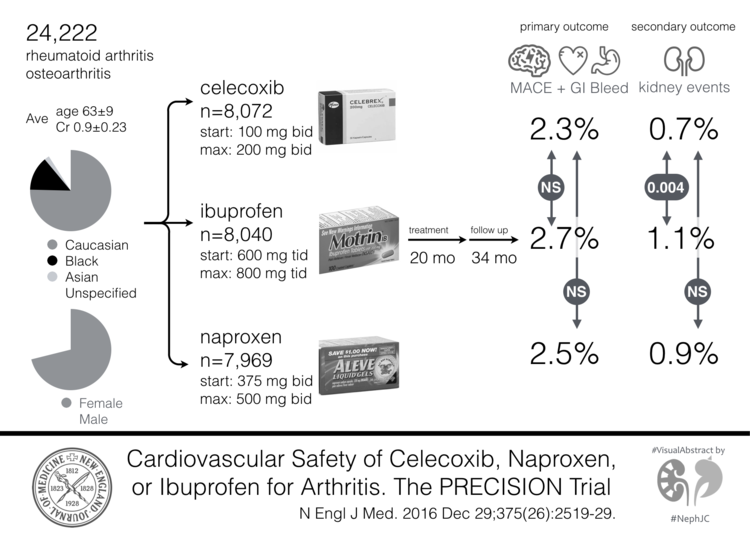
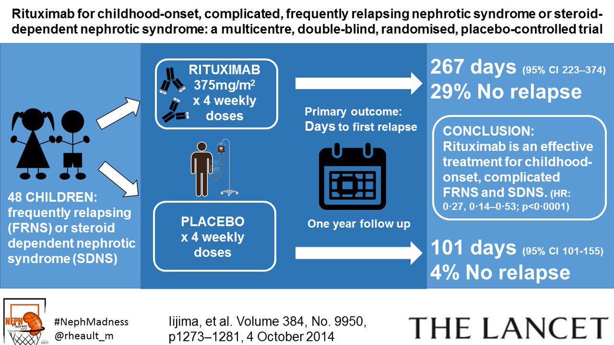
Following that, the #nephmadness 2017 has featured many visual abstracts, some pasted below


To top it all, CJASN is the first renal journal to enter in the visual abstract world. Great start and totally amazing to have a renal journal embrace this!


Here is an image showing early leaders of visual abstracts ( courtesy Andrew)


Few that I have done


How to create them? Check  out this primer by Andrew
You can create both static and animated abstracts- both can be very important in relaying your message.  I would urge all educators to try this out.

This could make fellowship journal club more fascinating. Make your residents/fellows create them. All researchers should simplify their ideas using such techniques.  All medicine journals should strongly consider this modality.

A new wave of presenting research has arrived- #visual abstracts!

Thursday, March 9, 2017

Concept Map: HSCT related renal disease: big picture


Here is a big picture overview of AKI and CKD seen with bone marrow transplant patients
Click on image for bigger picture.

AMACING trial: Fluids vs No fluids for Contrast Nephropathy prevention: A visualabstract


An animated #visualabstract for the recent AMACING study published in Lancet 2017 that was a randomized controlled trial for prevention of CIN with IV fluids vs placebo.

Below is a non animated version of the abstract


Monday, March 6, 2017

Nephmadness 2017: A Historical Perspective

NephMadness 2017 full logo

Every March, the nephrology social media scene is taken over by the one and the only- Nephmadness, an homage to the annual US college basketball tournament fondly known as March Madness. 

In early 2013, while I led the AJKD blog as editor, Joel Topf and Matt Sparks came up to me and said " Let's do March Madness version of Nephrology",  Being surrounded mostly by Cricket and soccer( the real football) most of my childhood and life, March Madness was something I learnt from my nephrology buddies. The idea was totally amazing!  What was even more amazing was that the AJKD editorial team took the risk with us on embarking on this venture.  And then it was born- March 2013- the first ever Nephmadness competition( the modern version of Nephrology Uptodate).  A collection of ideas, concepts competing year after year. 

I would recommend all to read prior years competitions- It's like reading a nephrology textbook but with fun and flavor and attitude-- totally cool! You can also read last year’s helpful guide, NephMadness for Dummies, for more details.

Prior years winners were


An editorial in AJKD highlighted this event in 2015. This is an example of reverse blogging leading to publication. Usually publications lead to blog posts and twitter events. The Nephmadness phenomenon lead to an invited editorial in the home journal describing the big event in Nephrology. The link is below

https://www.ncbi.nlm.nih.gov/pubmed/25704043

This year the tournament has 32 nephrology concepts divided across 8 different topic areas, called regions. Each region has 4 concepts. We have selected content experts from each topic to help us determine the best concepts and vet the information we provide to make sure it is accurate, unbiased, and interesting. The topics are secret until the contest begins on March 7, 2017- this year. Visit the AJKDBlog to get more information in 2 days.  


From 2013, to now, we have come a long way. More participants, more faculty, more involvement. This might have become the largest Medical online Game in history-- probably! I don't know of anything like this. If you do, let me know. If you don't, let the World records holders be ware!

Come play and enjoy! and most important of all- learn.
Students, fellows, residents, attendings, fellowship programs, all can participate.
Check out @kidneyCathy from Australia doing a "what is nephmadness" for all of us. 


Follow the NephMadness Team (listed below) to get engaged!